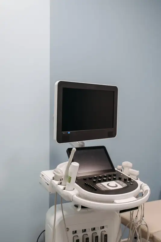
Recent advancements in wearable technology have led to the development of a groundbreaking device that utilizes ultrasound to assess the stiffness of human tissue. This innovative approach has the potential to enhance the detection and management of various medical conditions, track lesion progression, and evaluate rehabilitation outcomes.
Engineers at the University of California San Diego have designed a stretchable ultrasonic array capable of performing non-invasive, three-dimensional imaging of tissues located up to 4 cm beneath the skin’s surface, achieving a spatial resolution of 0.5 mm. This novel technique provides a longer-term and non-invasive alternative to existing methods, significantly improving penetration depth.
The research, led by Sheng Xu, a nanoengineering professor at UC San Diego, has resulted in a wearable device that continuously evaluates tissue stiffness. By integrating an array of ultrasound elements within a soft elastomer matrix and employing wavy serpentine stretchable electrodes, the device can conform to the skin, allowing for serial assessments of tissue stiffness.
According to Hongjie Hu, a postdoctoral researcher and co-author of the study, this elastography monitoring system can offer continuous, non-invasive, and three-dimensional mapping of mechanical properties in deep tissues. The implications of this technology are vast:
- In medical research, it can provide critical data on the progression of diseases such as cancer, which often leads to tissue hardening.
- It can assist in diagnosing and treating sports injuries by monitoring muscles, tendons, and ligaments.
- For cardiovascular and liver diseases, as well as certain chemotherapy treatments, continuous elastography can evaluate tissue stiffness and treatment efficacy, potentially leading to new therapeutic strategies.
Beyond cancer monitoring, this technology has additional applications:
- It can track liver cirrhosis and fibrosis, enabling healthcare professionals to monitor disease progression and tailor treatment plans effectively.
- It can evaluate musculoskeletal disorders such as tennis elbow and carpal tunnel syndrome, providing insights into the progression of these conditions for personalized treatment.
- It can aid in diagnosing myocardial ischemia by monitoring arterial wall elasticity, allowing for early intervention to prevent further damage.
The Xu Lab: Pioneering Wearable Ultrasound Technology
Over the years, the Xu lab has emerged as a leader in wearable ultrasound technology, transforming traditional portable devices into stretchable, wearable solutions. This innovation allows for continuous health monitoring, breaking the limitations of conventional ultrasound methods, which often require hospital settings and professional operation.
Hu emphasizes that this technology enables patients to monitor their health status anytime and anywhere, potentially reducing misdiagnoses and healthcare costs by providing a non-invasive and affordable alternative to traditional diagnostic procedures.
The rise of wearable ultrasound technology is revolutionizing healthcare monitoring, enhancing patient outcomes, and promoting the adoption of point-of-care diagnostics. As this technology evolves, significant advancements in medical imaging and healthcare monitoring are anticipated.
Yuxiang Ma, another co-author of the study, highlights the device’s ability to adapt to human skin and acoustically couple with it, facilitating precise elastographic imaging validated by magnetic resonance elastography.
Operational Mechanism
The device employs a 16 by 16 array of ultrasound elements, each constructed from a 1-3 composite and a backing layer made from a silver-epoxy composite to absorb excess vibrations, enhancing axial resolution. The chosen center frequency of 3 MHz strikes a balance between high spatial resolution and effective tissue penetration.
During testing, the device was utilized to map the three-dimensional distribution of Young’s modulus in tissues, detecting microstructural damage in muscles before soreness onset and tracking recovery during physiotherapy.
Specifications
- Dimensions: approximately 23 mm × 20 mm × 0.8 mm
- Biaxial stretchability: 40%
- Penetration depth: over 4 cm
- Highest signal-to-noise ratio: 28.4 dB
- Spatial resolution: 0.5 mm
- Contrast resolution: 1.74 dB
Addressing Challenges
To capture the motion of scattering particles and assess their displacement fields, the technology must record weak reflected signals, which requires sensitive technology. Traditional fabrication methods can cause thermal damage to piezoelectric materials, degrading transducer sensitivity.
To overcome these challenges, the researchers developed a low-temperature bonding approach using conductive epoxy, allowing for room temperature bonding without damaging the elements. Additionally, they replaced the single plane wave transmission mode with a coherent plane-wave compounding mode, enhancing signal intensity across the sample.
Future Directions
Future enhancements may include integrating a calibration layer with known modulus to obtain quantitative values of tissue moduli, further improving diagnostic capabilities. Advanced fabrication techniques could also refine the array design, increasing spatial resolution and expanding the sonographic window.
Gao notes the potential for collaboration with physicians to explore practical applications in clinical settings, emphasizing the device’s promise for monitoring high-risk groups and enabling timely interventions.
